
Keck researchers use bioluminescence to treat cancer
To develop new ways to improve cancer treatments in humans, researchers at the Keck School of Medicine recently studied the enzymes in marine animals that cause bioluminescence.
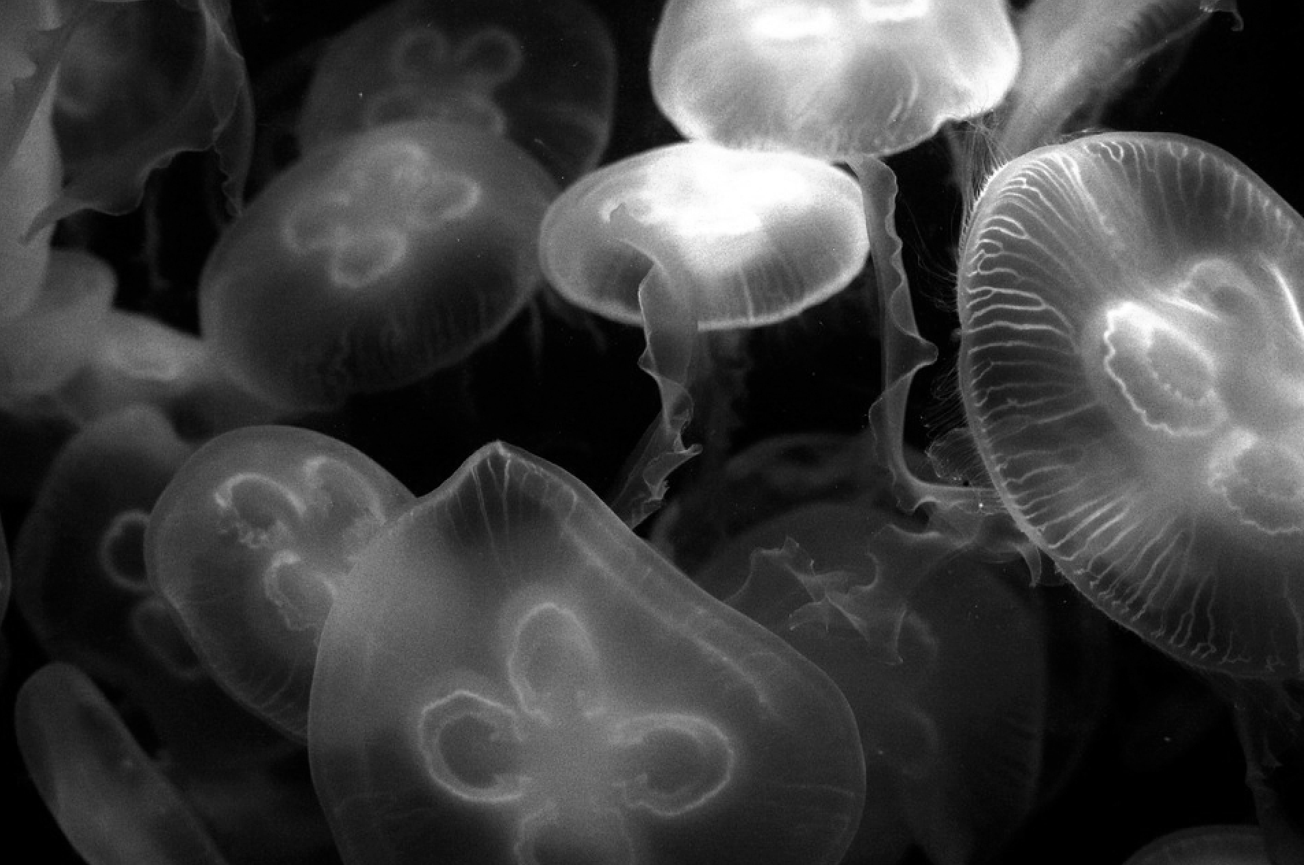
Marine animals with bioluminescence, like the jellyfish pictured above, are helping a team of researchers at the Keck School of Medicine develop effective immunotherapy cancer treatments. Photo from Pixabay.
The team of researchers utilized these enzymes to develop a test to determine whether immunotherapies — uses of the immune system to fight cancer — are effective, according to a Scientific Reports study published on Jan. 9. The bioluminescent property of these enzymes allowed the researchers to see if a therapy is successful in killing cancer cells.
“[Immunotherapy] can use the body’s immune system to fight against several diseases including cancer in a similar way the body fights against foreign invaders,” said Hittu Matta, a Keck assistant professor of research medicine who contributed to the study.
According to Matta, a number of immunotherapies such as chimeric antigen receptor-T cells (CAR-T) have been developed. Chimeric antigen receptors are proteins that help T cells recognize toxins on targeted cancer cells.
However, since these are developed for a specific patient and not mass produced, creating them is expensive and having an accurate test to ensure their effectiveness is significant.
As a fairly new advancement in the cancer research field, immunotherapies can help scientists determine the next steps in developing proper cancer treatments, Matta said. This can be achieved through testing the therapies quickly, safely and cheaply.
“Cancer immunotherapies are one of the hottest areas of cancer research,” said Keck professor Preet Chaudhary, the corresponding author of the study. “To develop more effective and less toxic immunotherapies, it is important to have an assay that can rapidly screen a large number of candidates in a sensitive, fast, economical manner to select the best candidate for further development.”
Named after the El-Matador Beach, the Matador assay makes use of bioluminescent agents to visually display and measure how many cancer cells are being killed by a selected immunotherapy.
“The [agents were] engineered to stay entrapped inside the intact cells, but it gets released after the cells die, and the glow released from them was quantitatively measured using a luminometer,” Matta said.
According to Matta, the technology used in the Matador assay has the potential to impact several fields outside of medicine, including light production, genetically engineered plants and environmental technology.