Imaging tool improves bacteria detection
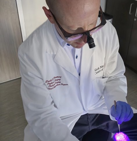
Autofluorescence imaging can lead bacteria in wounds to glow visible red or cyan.
Autofluorescence imaging can lead bacteria in wounds to glow visible red or cyan.

New applications of biomedical imaging via autofluorescence tools can visually “light up” bacteria, allowing clinicians to debride — or remove non-viable tissue in wounds — and prevent infection, according to a research study published by Keck Medicine of USC in “Advances in Wound Care.”
Some health conditions, including diabetes, can increase the risk of developing chronic wounds, also known as non-healing wounds. About half of diabetic foot ulcers become infected — and an estimated 20% can require amputations.
Chronic wounds, which occur when tissue does not heal within an expected time frame, can become susceptible to bacterial growth and infections. Wound infections can require treatment with antibiotics or, in critical cases, amputation.
Certain health conditions can affect healing processes due to underlying disease pathophysiology. Diabetic neuropathy — damage to the nerves — can alter sensations of pain and predispose patients to diabetic foot ulcers and severe infections.
Infections, while sometimes visible by signs of inflammation or blistering skin, are not indicative of the extent of bacterial load in wounds. Current clinical diagnostic methods, such as sampling and assessing bacteria, can assist in infection treatment. Detecting bacteria more accurately and efficiently allows clinicians to remove necrotic tissue and promote healing of a wound.
The study evaluated the use of autofluorescence imaging, which can be used to make bacteria “glow” by transillumination.
“If we see a lot of bacteria in an area, then that might be someone that might be at risk for getting a wound,” said Dr. David Armstrong, a podiatric surgeon and limb preservation specialist with Keck Medicine of USC, who led the research study. “We can debride that away gently and efficiently and maybe stop the infection before it starts.”
Clinicians assessing the extent of infection may be able to use the AF imaging tool to visualize the location of bacteria present in non-healing wounds. The technology can be affixed to a handheld or to a head-mounted display, Armstrong said.
AF imaging is used in other biomedical applications to assist in the diagnosis of retinal disease by detecting the presence of fluorophores — or molecules that light up under microscope imaging — in the eye.
In infection prevention, the innovation is promising: Nearly 90% of bacteria present in chronic wounds display AF properties. Clinical providers can use AF to make bacteria glow red or cyan, which allows for more efficient debriding of wounds.
“Since technology can identify indicators of things that could get worse, [it] could help prevent things before the condition exacerbates,” said Shreeya Chand, a freshman majoring in computer science.
Roshni Lulla, a graduate student studying psychology, said the health advancements could improve early diagnostics and benefit health outcomes.
“Technology has a huge impact, especially [with] the rise of [artificial intelligence] recently, you can detect things a lot quicker [and] can help diagnose,” Lulla said. “Especially [for] someone who has diabetes or any other comorbidities, they have so many other things that they have to think about [and] there’s additional risk of having infections.”
A new diabetes-associated wound occurs every second globally, Armstrong said.
Other underlying health issues and conditions — including poor circulation, weakened immune system functioning and chronic kidney disease — can also increase the risk of non-healing wounds, according to other studies.
Armstrong’s study, a literature review of 25 studies, considers the application of preexisting biomedical technology to new tools — and the impact in advancing diagnostics and prevention of chronic wounds, severe infections and amputations.
Armstrong hopes the AF imaging will improve health outcomes for patients.
“This may be one of those kinds of instruments that might help us better measure what we manage,” Armstrong said. “If we can identify these problems, like infections, and we can get in front of them by being able to identify these risk factors, then maybe we can make a difference.”
Editor’s Note: A previous version of this article misrepresented Dr. David Armstrong’s affiliation to the University. He works for Keck Medicine of USC, not Keck School of Medicine of USC. The article was updated Sept. 15 at 4 p.m. to reflect this change.
Editor’s Note: A previous version of this article stated Dr. David Armstrong worked for Keck Medicine of USC and not Keck School of Medicine of USC. He works at both entities, but his study was a part of his clinical work done under Keck Medicine of USC. The article was updated Sept. 23 at 3 p.m. to reflect this change.
We are the only independent newspaper here at USC, run at every level by students. That means we aren’t tied down by any other interests but those of readers like you: the students, faculty, staff and South Central residents that together make up the USC community.
Independence is a double-edged sword: We have a unique lens into the University’s actions and policies, and can hold powerful figures accountable when others cannot. But that also means our budget is severely limited. We’re already spread thin as we compensate the writers, photographers, artists, designers and editors whose incredible work you see in our daily paper; as we work to revamp and expand our digital presence, we now have additional staff making podcasts, videos, webpages, our first ever magazine and social media content, who are at risk of being unable to receive the support they deserve.
We are therefore indebted to readers like you, who, by supporting us, help keep our paper daily (we are the only remaining college paper on the West Coast that prints every single weekday), independent, free and widely accessible.
Please consider supporting us. Even $1 goes a long way in supporting our work; if you are able, you can also support us with monthly, or even annual, donations. Thank you.
This site uses cookies. By continuing to browse the site, you are agreeing to our use of cookies.
Accept settingsDo Not AcceptWe may request cookies to be set on your device. We use cookies to let us know when you visit our websites, how you interact with us, to enrich your user experience, and to customize your relationship with our website.
Click on the different category headings to find out more. You can also change some of your preferences. Note that blocking some types of cookies may impact your experience on our websites and the services we are able to offer.
These cookies are strictly necessary to provide you with services available through our website and to use some of its features.
Because these cookies are strictly necessary to deliver the website, refusing them will have impact how our site functions. You always can block or delete cookies by changing your browser settings and force blocking all cookies on this website. But this will always prompt you to accept/refuse cookies when revisiting our site.
We fully respect if you want to refuse cookies but to avoid asking you again and again kindly allow us to store a cookie for that. You are free to opt out any time or opt in for other cookies to get a better experience. If you refuse cookies we will remove all set cookies in our domain.
We provide you with a list of stored cookies on your computer in our domain so you can check what we stored. Due to security reasons we are not able to show or modify cookies from other domains. You can check these in your browser security settings.
These cookies collect information that is used either in aggregate form to help us understand how our website is being used or how effective our marketing campaigns are, or to help us customize our website and application for you in order to enhance your experience.
If you do not want that we track your visit to our site you can disable tracking in your browser here:
We also use different external services like Google Webfonts, Google Maps, and external Video providers. Since these providers may collect personal data like your IP address we allow you to block them here. Please be aware that this might heavily reduce the functionality and appearance of our site. Changes will take effect once you reload the page.
Google Webfont Settings:
Google Map Settings:
Google reCaptcha Settings:
Vimeo and Youtube video embeds:
The following cookies are also needed - You can choose if you want to allow them:
